

Ready-To-Use 3D Human Retinal Pigment Epithelial Spheroids
Product Code:
SC-SP3D-6540
SC-SP3D-6540
Host Type:
Human
Human
Regulatory Status:
RUO
RUO
Shipping:
Dry Ice
Dry Ice

No additional charges, what you see is what you pay! *
| Code | Size | Price |
|---|
| SC-SP3D-6540 | 1 kit | £1,263.00 | |||||||||||||||||||||||||||||||||||||||||||||||||||||||||||||||||||||||||||||||||||||||||||||||||
| Available in 24, 48 or 96 plates. Contact us for pricing. | |||||||||||||||||||||||||||||||||||||||||||||||||||||||||||||||||||||||||||||||||||||||||||||||||||
Quantity:
Prices exclude any Taxes / VAT
Stay in control of your spending. These prices have no additional charges, not even shipping!
* Rare exceptions are clearly labelled (only 0.14% of items!).
* Rare exceptions are clearly labelled (only 0.14% of items!).
Multibuy discounts available! Contact us to find what you can save.
This product comes from: United States.
Typical lead time: 10-14 working days.
Contact us for more accurate information.
Typical lead time: 10-14 working days.
Contact us for more accurate information.
- Further Information
- Documents
- Related Products
- Show All
Further Information
Description:
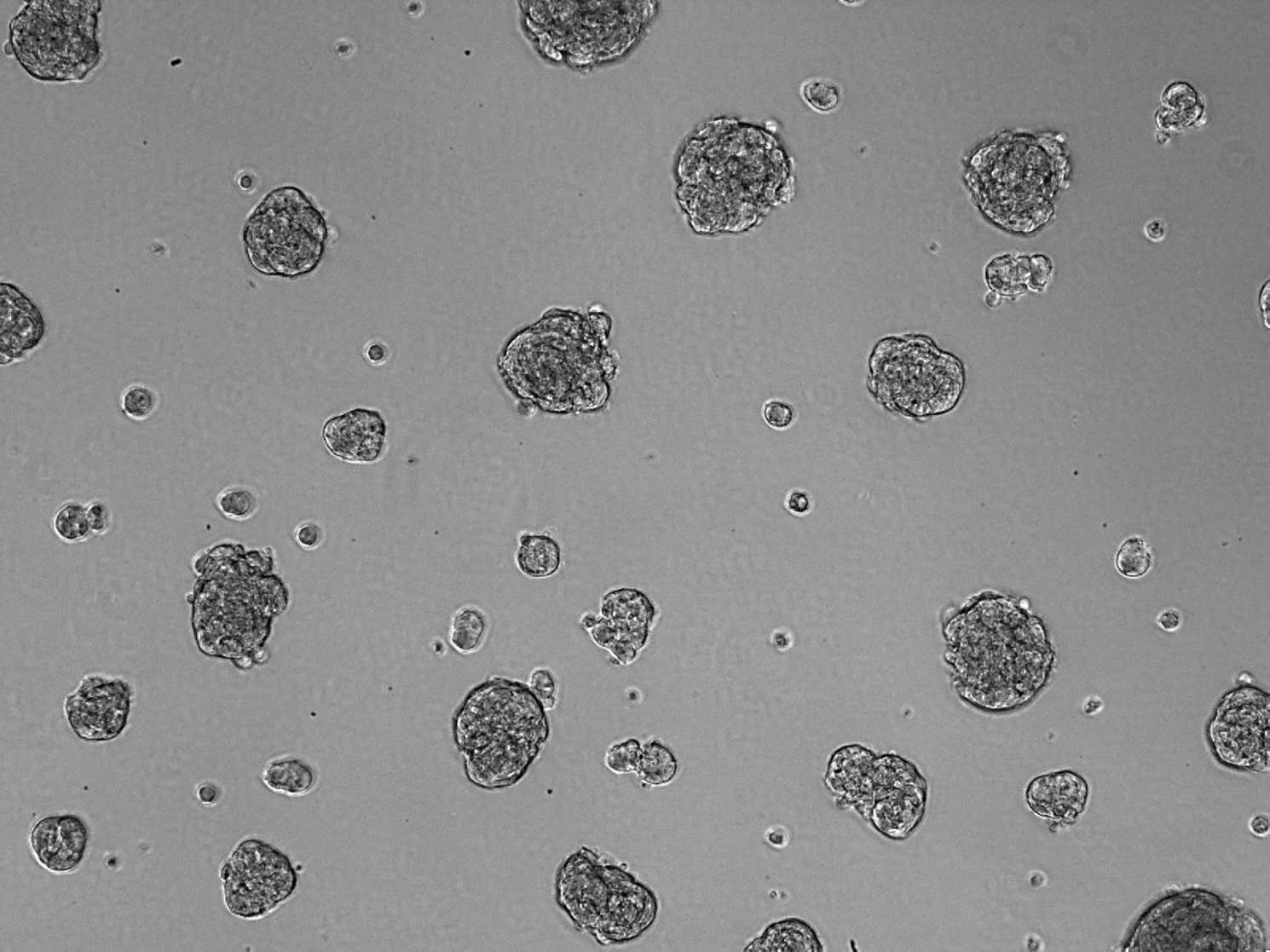
Age-related macular degeneration (AMD) is characterized in its early stages by the presence of extracellular deposits, known as drusen, that accumulate between the basal surface of the retinal pigmented epithelium and Bruch's membrane, an extracellular matrix complex that separates the neural retina from the capillary network in the choroid [1]. Several studies have shown that drusen contains a variety of protein and lipid components [2]. Although liver is the primary biosynthetic site for most of these molecules, retinal pigment epithelial (RPE) cells locally synthesize a number of drusen components [2]. The respective contributions of RPE-derived and plasma-derived molecules to the biogenesis of drusen, and the relevant molecular interactions leading to drusen depositions, however, have not been fully identified. One of the major limitations is that RPE cells, once isolated from the eye, tend to dedifferentiate into myofibroblasts in conventional 2D cell culture. Recent studies have shown that 3D retinal pigment epithelial cell spheroids form and maintain a well-differentiated epithelium in 3D cell culture [3]. ScienCell Research Laboratories, as a result, has developed ready-to-use 3D human retinal pigment epithelial spheroids (SP3D-HRPEpiS). 3D RPE spheroids exhibit the differentiated epithelial cell marker cytokeratin-18 and deposit apolipoprotein ApoE, a prominent drusen constituent. The 3D RPE spheroid model is an ideal way to model drusen in vitro and study the pathogenesis of related diseases, such as AMD.
Kit Components:
3D Cell Culture Components
- 1 x #SP-6540 - Human Retinal Pigment Epithelial Cells (SP-HRPEpiC) - 1 x 10^4 speroids - Storage: Liquid nitrogen
- 1 x #3D-4101 - 3D-Epithelial Spheroid Medium (3D-EpiSpM) - 200 mL - Storage: 2-8°C
- 1 x #0004 - Fetal Bovine Serum (FBS) - 4 mL - Storage: -20°C
- 1 x #0583 - Penicillin/Streptomycin Solution (P/S) - 2 mL - Storage: -20°C
- 1 x #0343 or #0353 or #0383 - Ultra-Low Binding Culture Plates (24-, 48-, or 96- well plate) - 1 plate - Storage: RT
Extra Description:
ScienCell's 3D human retinal pigment epithelial spheroids display the differentiated epithelial cell marker cytokeratin-18 and deposit apolipoprotein ApoE, a prominent drusen constituent. The 3D RPE spheroid model is an ideal way to model drusen in vitro and study the pathogenesis of related diseases, such as AMD.
Documents
Related Products
| Product Name | Product Code | Supplier | All-inclusive 3D Human Retinal Pigment Epithelial Spheroid Formation Kit | SC-3D-6540 | Sciencell | Summary Details | |||||||||||||||||||||||||||||||||||||||||||||||||||||||||||||||||||||||||||||||||||||||||||||
|---|---|---|---|---|---|---|---|---|---|---|---|---|---|---|---|---|---|---|---|---|---|---|---|---|---|---|---|---|---|---|---|---|---|---|---|---|---|---|---|---|---|---|---|---|---|---|---|---|---|---|---|---|---|---|---|---|---|---|---|---|---|---|---|---|---|---|---|---|---|---|---|---|---|---|---|---|---|---|---|---|---|---|---|---|---|---|---|---|---|---|---|---|---|---|---|---|---|---|---|