ProSci

PDL1 Antibody [1D7]

Product Code:
 
PSI-RF16038
Product Group:
 
Primary Antibodies
Supplier:
 
ProSci
Host Type:
 
Mouse
Antibody Isotype:
 
IgG1
Antibody Clonality:
 
Monoclonal
Antibody Clone:
 
1D7
Regulatory Status:
 
RUO
Target Species:
 
Human
Applications:
  • Enzyme-Linked Immunosorbent Assay (ELISA)
  • Immunocytochemistry (ICC)
  • Immunofluorescence (IF)
  • Immunohistochemistry- Paraffin Embedded (IHC-P)
  • Western Blot (WB)
Shipping:
 
blue ice
Storage:
 
PD-L1 antibody can be stored at 4˚C for three months and -20˚C, stable for up to one year. As with all antibodies care should be taken to avoid repeated freeze thaw cycles. Antibodies should not be exposed to prolonged high temperatures.
1 / 8
Western blot analysis of PD-L1 in overexpressing HEK293 cells PD-L1 antibody at 0.25 and 0.5 μg/ml
2 / 8
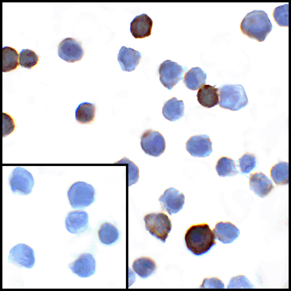
Immunocytochemistry of PD-L1 in transfected HEK293 cells with PD-L1 antibody at 1 μg/mL. Lower left: Immunocytochemistry in transfected HEK293 cells with control mouse IgG antibody at 1 μg/mL.
3 / 8
Immunofluorescence of PD-L1 in transfected HEK293 cells with PD-L1 antibody at 2 μg/mL. <br><br>Red: PDL1 Antibody [1D7] (RF16038) <br> Blue: DAPI staining
4 / 8
Immunofluorescence of PD-L1 in human stomach carcinoma tissue with PD-L1 antibody at 2 μg/mL. <br><br>Red: PDL1 Antibody [1D7] (RF16038) <br> Blue: DAPI staining
5 / 8
Immunofluorescence of PD-L1 in human tonsil tissue with PD-L1 antibody at 2 μg/mL. <br><br>Red: PDL1 Antibody [1D7] (RF16038) <br> Blue: DAPI staining
6 / 8
Immunohistochemistry of PD-L1 in human stomach carcinoma tissue with PD-L1 antibody at 5 μg/mL.
7 / 8
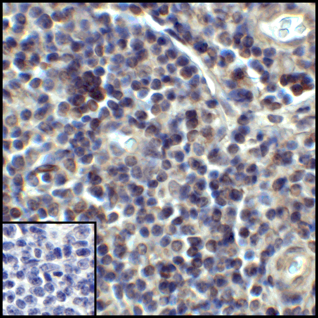
Immunohistochemistry of PD-L1 in human tonsil tissue with PD-L1 antibody at 5 μg/mL.
8 / 8
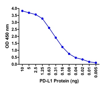
A sandwich ELISA was performed using the anti-PD-L1 mAb RF16038 (5 μg/ml) as the capture antibody. Biotin-labeled anti-PD-L1 mAb RF16032-biotin (1 μg/ml) and streptavidin-HRP (0.1 μg/ml) were used for detection. Detection range is from 10 ng to 20 pg.

Western blot analysis of PD-L1 in overexpressing HEK293 cells PD-L1 antibody at 0.25 and 0.5 μg/ml
Immunocytochemistry of PD-L1 in transfected HEK293 cells with PD-L1 antibody at 1 μg/mL. Lower left: Immunocytochemistry in transfected HEK293 cells with control mouse IgG antibody at 1 μg/mL.
Immunofluorescence of PD-L1 in transfected HEK293 cells with PD-L1 antibody at 2 μg/mL. <br><br>Red: PDL1 Antibody [1D7] (RF16038) <br> Blue: DAPI staining
Immunofluorescence of PD-L1 in human stomach carcinoma tissue with PD-L1 antibody at 2 μg/mL. <br><br>Red: PDL1 Antibody [1D7] (RF16038) <br> Blue: DAPI staining
Immunofluorescence of PD-L1 in human tonsil tissue with PD-L1 antibody at 2 μg/mL. <br><br>Red: PDL1 Antibody [1D7] (RF16038) <br> Blue: DAPI staining
Immunohistochemistry of PD-L1 in human stomach carcinoma tissue with PD-L1 antibody at 5 μg/mL.
Immunohistochemistry of PD-L1 in human tonsil tissue with PD-L1 antibody at 5 μg/mL.
A sandwich ELISA was performed using the anti-PD-L1 mAb RF16038 (5 μg/ml) as the capture antibody. Biotin-labeled anti-PD-L1 mAb RF16032-biotin (1 μg/ml) and streptavidin-HRP (0.1 μg/ml) were used for detection. Detection range is from 10 ng to 20 pg.

No additional charges, what you see is what you pay! *

CodeSizePrice
PSI-RF16038-0.02mg0.02mg£150.00
Quantity:
PSI-RF16038-0.1mg0.1mg£515.00
Quantity:
Prices exclude any Taxes / VAT
Stay in control of your spending. These prices have no additional charges, not even shipping!
* Rare exceptions are clearly labelled (only 0.14% of items!).
Multibuy discounts available! Contact us to find what you can save.
This product comes from: United States.
Typical lead time: 14-21 working days.
Contact us for more accurate information.
  • Further Information
  • Documents
  • Show All

Further Information

Additional Names:
PD-L1 Antibody: Programmed cell death 1 ligand-1, programmed death ligand 1, PDL1, PDL-1, B7-H1
Application Note:
PD-L1 antibody can be used for detection of PD-L1 by Western blot at 0.5 - 1 μg/mL. Antibody can also be used for immunohistochemistry starting at 2 - 5 μg/mL. For immunofluorescence start at 5μg/mL.

Antibody validated: Western Blot in human samples; Immunohistochemistry in human samples; Immunocytochemistry in human samples and Immunofluorescence in human samples. All other applications and species not yet tested.
Background:
PD-L1 Antibody: Cell-mediated immune responses are initiated by T lymphocytes that are themselves stimulated by cognate peptides bound to MHC molecules on antigen-presenting cells (APC) (1). T-cell activation is generally self-limited as activated T cells express receptors such as PD-1 (also known as PDCD-1) that mediate inhibitory signals from the APC (2). PD-1 can bind two different but related ligands, PD-L1 and PD-L2. PD-L1 is a B7-related protein that inhibits cell-mediated immune responses by reducing the secretion of IL-2 and IL-10 from memory T cells (3). This suggests that PD-L1 may be useful in reducing allogenic CD4+ memory T-cell responses to endothelial cells, thereby reducing the likelihood of host immune responses to allografts. PD-L1 also functions as an immune checkpoint protein, and multiple anti-PD-L1 antibodies are currently in phase II and III clinical trials, with one antibody already approved for the treatment of cancer (4).
Background References:
  • Holling TM, Schooten E, and van Den Elsing PJ. Function and regulation of MHC class II molecules in T-lymphocytes: of mice and men. Hum. Immunol. 2004; 65:282-90.
  • Ishida Y, Agata Y, Shibahara K, et al. Induced expression of PD-1, a novel member of the immunoglobulin gene superfamily, upon programmed cell death. EMBO J. 1992; 11:3887-95.
  • LaGier J and Pober JS. Immune accessory functions of human endothelial cells are modulated by overexpression of B7-H1 (PDL1). Hum. Immunol. 2006; 67:568-78.
  • Aydin AM, Woldu SL, Hutchinson RC, et al. Spotlight on atezolizumab and its potential in the treatment of advanced urothelial bladder cancer.Onco. Targets Ther. 2017;10:1487-502.
Buffer:
PD-L1 Antibody is supplied in PBS containing 0.02% sodium azide and 50% glycerol.
Concentration:
1 mg/mL
Conjugate:
Unconjugated
DISCLAIMER:
Optimal dilutions/concentrations should be determined by the end user. The information provided is a guideline for product use. This product is for research use only.
Immunogen:
PD-L1 antibody was raised against the extracellular domain of human PD-L1.
NCBI Gene ID #:
29126
NCBI Official Name:
programmed cell death 1 ligand
NCBI Official Symbol:
CD274
NCBI Organism:
Homo sapiens
Physical State:
Liquid
PREDICTED MOLECULAR WEIGHT:
Predicted: 32 kDa

Observed: 45 kDa
Protein Accession #:
NP_054862
Protein GI Number:
7661534
Purification:
PD-L1 Antibody is supplied as protein A purified IgG1.
Research Area:
Immunology
Swissprot #:
Q9NZQ7
User NOte:
Optimal dilutions for each application to be determined by the researcher.