Drug Discovery Tools
Cells consist of various elements such as nucleic acid, proteins and substructures. Moreover, functions are dynamically regulated by signal transduction. Purified protein can provide some observation in drug discovery but the biological relevance of this information is not always clear.
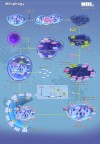
In order to overcome this challenge, MBL International Corporation offers fluorescent protein-based drug discovery tools in living cells. Their novel technology, Fluoppi, can visualize Protein-Protein Interaction (PPI) of interest in living cells. Moreover, other product offering such as Keima-Red and FUCCI, can visualise autophagy/mitophagy and cell cycle respectively. These technologies are applicable for cell-based high-throughput drug screening.
Fluoppi: Protein-Protein Interaction Detection in Living Cells
Fluoppi (Fluorescent based technology detecting Protein-Protein Interaction) is a novel technology to detect Protein-Protein Interaction (PPI) in living cells with a high signal to noise ratio. The result is a bright, clearly analysable image. Fluoppi detects PPI as the absence or presence of fluorescent foci during inhibition or induction, respectively. The Fluoppi advantage is the ease of the construction of the PPI detection system with no need for optimising the linker.

PPI Induction
PPI Inhibition
FUCCI: Fluorescent Ubiquitination-based Cell Cycle Indicator (FUCCI)
FUCCI (Fluorescent Ubiquitination-based Cell Cycle Indicator) is a set of fluorescent probes which enables the visualisation of cell cycle progression in living cells. FUCCI utilises the phase-dependent nature of replication licensing factors Cdt1 and Geminin. A fusion protein of a fragment of Cdt1 (amino acids 30-120) with the fluorescent protein monomeric Kusabira-Orange 2 (mKO2) serves as an indicator of G1 phase. A fusion protein of a fragment of Geminin (amino acids 1-110 or 1-60) with the fluorescent protein monomeric Azami-Green 1 (mAG1) visualises the S,G2 and M phase. In summary, FUCCI utilises the highly selective, rapid degradation of the replication licensing factors mediated by the ubiquitin proteasome system to give excellent visualisations of the cell cycle.

What makes FUCCI a powerful research tool?
- Real-time visualisation of cell cycle progression
- Spatio-temporal imaging of cell cycle dynamics
- Utilisation of fluorescent proteins Green mAG1and Orange mKO2
- Fucci-G1 Orange labels G1 phase nuclei in orange. Fucci-S/G2/M Green labels S/G2/M phases nuclei in green.
By visualising the cell cycle, Fucci is a powerful tool to investigate any process that has to do with cell growth and differentiation, such as the development and regeneration of organs as well as carcinogenesis.
Keima-Red for mitophagy detection in living cells
Mitophagy is the selective degradation of old or depolarised mitochondria by autophagy and contributes to maintaining a healthy population of mitochondria. Since damaged mitochondria lead to collapse cell homoeostasis, mitophagy is believed to be protective against diseases related to mitochondrial dysfunction such as in neurodegenerative disorders.
The fluorescent protein Keima has an excitation spectrum that changes according to pH. A short wavelength (440 nm) is predominant for excitation in a neutral environment, whereas a long wavelength (586 nm) is predominant in an acidic environment. The ratio of fluorescent intensity in each excitation condition is an indicator of mitophagy in living cells.


